
Breast cancer is the most common form of cancer among New Zealand
women. One in nine New Zealand women will develop breast cancer at some
stage of their lives.
Initial symptoms will normally include a new lump in the breast;
treatment will generally involve surgery followed by a combination of
other treatments such as chemotherapy or radiotherapy.
General information
In New Zealand breast cancer was the second most common cancer in
2012 and was the third leading cause of cancer death. In real numbers,
that translates to 3054 diagnoses and 618 deaths form the disease..
Among women breast cancer was the most common cancer (3025 diagnoses)
and the third most common cause of death (617 cases) behind lung cancer
and colorectal cancer.Around 30 men are diagnosed with the condition in New Zealand each year which equates to around 1% of all breast cancer diagnoses.
Breast cancer occurs when breast cells develop abnormally and grow
out of control forming a malignant (cancerous) tumour. It is possible
for cancer cells to spread (metastasise) from the breast to other parts
of the body via the lymphatic system and by direct entry into the blood
stream. Once the cancer cells have reached other body sites they can
form "secondary" cancers.
Prior to menopause the
majority of new breast lumps found are benign (non-cancerous).However,
after menopause one in two new breast lumps found will be malignant.
Approximately 75% of all breast cancers will occur in women over the age
of 50 years.
Breast cancer can recur. Once a woman has had breast cancer her chances of developing it again are increased five-fold.
About breasts
Breasts are composed of fibrous (connective) tissue, fatty tissue and
glandular tissue. They lie on a band of strong muscle that sits on the
ribs of the chest. Breasts have no useful biological function other than
to provide milk for babies.Fibrous tissue gives the breast its shape and helps to support the fatty tissue and glandular tissue. As women age, the fibrous tissue is replaced with fatty tissue. This leads to changes in the shape and texture of the breasts.
The glandular tissue is made up of a series of lobules. These are small structures that produce milk when stimulated by female hormones during pregnancy. The milk produced in the lobules drains into small channels or ducts that eventually open into the nipple. Glandular tissue also responds to menstrual-related hormonal changes, which can cause the breasts to be occasionally tender and lumpy.
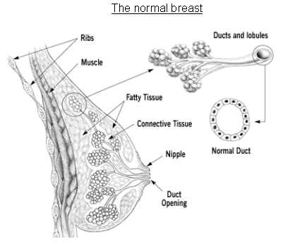
Picture reproduced with the kind permission of the Anti-Cancer Council of Victoria
In the breast, armpit and neck there are small glands called lymph
nodes, which, along with the lymph vessels into which the nodes drain,
are part of the lymphatic system. The role of the fluid filled lymphatic
system is to filter infections and prevent them entering the blood
stream. Each breast has a network of lymph vessels that drain into a
system of about 24 lymph nodes in the armpit. These are called the
axillary lymph nodes. There are also lymph nodes behind the breastbone
called the internal mammary nodes. Some of these nodes drain into the
axillary lymph nodes, with the remainder draining into other nodes in
the chest.
Axillary lymph nodes
Causes and risk factors
The causes of breast cancer are not known. However, it is known which women are more at risk of developing the condition. The main risk factors for developing breast cancer are:- Being a woman over the age of 40 years
- Having a family history of breast cancer - the younger the family member was when they developed breast cancer, the greater the risk
- Having had breast cancer previously
- Having had a biopsy showing an "at risk" breast lump or thickening
- Having a faulty gene, such as BRCA1 or BRCA2. Women with a BRCA1 gene mutation have a 55-65% risk of developing breast cancer by age 70 years. For women with a BRCA2 gene mutation the risk is approximately 45%.
- Having had an early onset of periods or the late onset of menopause
- Having had a first child after the age of 30 years or not having had children at all
- A diet high in fat, excessive alcohol and a reduced intake of fibre, fruits and vegetables
- Being on Hormone Replacement Therapy (HRT) medication for longer than 5-7 years
- Taking the oral contraceptive pill may slightly increase the risk of developing breast cancer, but this has not been conclusively proven
- Having dense breasts - dense breasts do not increase the risk of developing breast cancer, but they may make lumps difficult to feel and see.
Types of breast cancer
There are several different types of breast cancer. The two most
common types are ductal breast cancer (to do with the milk ducts) and
lobular breast cancer (to do with the milk lobules). Each of these
breast cancers can be either “in situ” (in the same place) or “invasive”
(has spread to normal tissue).
In situ carcinoma: These are pre-cancers and are the
earliest stage of breast cancer; they can either develop into invasive
breast cancer or raise the risk of developing invasive cancer. Caught
and treated early, they are often completely curable.
Ductal carcinoma in situ (DCIS): This is where
the breast cancer cells are completely contained within the milk ducts
and have not spread into the surrounding breast tissue. DCIS is usually
treated with surgery (mastectomy) or combined surgery (partial
mastectomy) and radiotherapy.
Lobular carcinoma in situ (LCIS): This is where
the breast cancer cells are completely contained within the milk lobules
and have not spread into the surrounding breast tissue. Often LCIS does
not need treatment. Instead, regular breast exams and mammograms may be
used to monitor for the early changes of developing breast cancer.
Invasive carcinoma: This is where the ductal or lobular
cancer spreads into the surrounding tissues. Approximately 90% of
invasive breast cancers are ductal cancers.
Other less common breast cancers include inflammatory breast cancer and medullary breast cancer.
Signs and symptoms
Most commonly, the first sign of breast cancer is a new lump in the breast. The lump is usually painless. Other signs of breast cancer include:- A new area of thickened tissue in the breast
- Nipple discharge or a change in the nipple
- Dimpling or puckering of the skin of the breast
- A change in breast size or shape.
While these symptoms may not be related to breast cancer, it is
important to see a doctor promptly for assessment and accurate diagnosis
if any of these symptoms are present. Early detection is vital in the
successful treatment of breast cancer.
Diagnosis
If an abnormal lump is found, or other symptoms are present, a
referral to a breast specialist for assessment and diagnosis will
probably be recommended. In order for an accurate diagnosis to be made
the three-step approach of clinical examination, imaging (mammography
and ultrasound scanning), and biopsy will be required.
Clinical examination:
The doctor will begin by examining both breasts. They will then check the abnormal lump's size and location, and other characteristics such as whether it is mobile, hard or soft, regular or irregular. The doctor will ask about the history of the lump such as how long has it been there, has it grown, is it painful. Risk factors such as family history or previous breast lumps will be discussed.
The doctor will begin by examining both breasts. They will then check the abnormal lump's size and location, and other characteristics such as whether it is mobile, hard or soft, regular or irregular. The doctor will ask about the history of the lump such as how long has it been there, has it grown, is it painful. Risk factors such as family history or previous breast lumps will be discussed.
Imaging (mammograms and / or ultrasound scanning):
A mammogram (specialised breast x-ray) shows the soft tissue of the breast and can indicate any suspicious areas. Ultrasound scanning uses sound waves to form an image of the breast tissue. Pictures of any suspicious areas can be taken. Ultrasound scanning is particularly useful for assessing whether a lump is fluid filled or solid.
A mammogram (specialised breast x-ray) shows the soft tissue of the breast and can indicate any suspicious areas. Ultrasound scanning uses sound waves to form an image of the breast tissue. Pictures of any suspicious areas can be taken. Ultrasound scanning is particularly useful for assessing whether a lump is fluid filled or solid.
There are different types of biopsies used to take cells or tissue samples from a suspicious lump so they can be sent to a laboratory for analysis under a microscope.
- Fine needle aspiration: This is usually the first type of biopsy used. It is performed using a local anaesthetic and involves inserting a fine needle into the lump and removing a small sample of cells and/or fluid. At the laboratory the sample is spread onto a glass slide and analysed. The insertion of the needle may be guided by ultrasound.
- Core biopsy: This uses a larger needle to remove a sample of tissue from the lump. A local anaesthetic is used and a very small incision (1-2mm) is made in the skin over the lump. The needle is usually guided into the lump by ultrasound. At the laboratory the tissue sample is sliced very finely and placed on a glass slide for analysis.
- Stereotactic core biopsy: This is a core biopsy performed on a special x-ray table allowing three-dimensional computerised images of the lump to be taken and used to guide the biopsy needle into the lump. This is useful for testing lumps seen on a mammogram that cannot be felt or visualised using an ultrasound scanner.
- Excision biopsy: This is a minor surgical procedure where part or all of the abnormal area is removed. It can be performed using a local or general anaesthetic. If the lump is unable to be precisely located using mammogram or ultrasound scanning, it may need to be marked by a thin wire called a "hookwire". This is inserted under x-ray guidance using a local anaesthetic just prior to the surgery.
If a diagnosis of breast cancer is made, blood tests, x-rays and
scans of the bones and liver may be performed to assess whether the
cancer has spread to other organs.
Stages of breast cancer
After diagnosis, breast cancers are assigned a "stage". The stage
indicates the tumour's size and how far it has spread within the breast,
surrounding tissues or to other organs in the body. Stages range from 0
to IV - a higher stage indicates more severe cancer.
Stage 0: The cancer has not spread beyond the ducts of the breast (ie: ductal carcinoma in situ or DCIS).
Stage I: These tumours measure less than two
centimetres. The axillary lymph nodes are not affected and there are no
signs that the cancer has spread elsewhere in the body.
Stage II: These tumours measure between two and
five centimetres, or the axillary lymph nodes are affected, or both.
There are no signs that the cancer has spread elsewhere in the body.
Stage III: These tumours are larger than five
centimetres, the axillary lymph nodes are usually affected, but there
are no signs that there has been any further spread.
Stage IV: These tumours are of any size, but the
axillary lymph nodes are usually affected and the cancer has spread to
other parts of the body.
Breast cancer tumours are also classified as being "hormone
receptor positive" or "hormone receptor negative". Approximately 60% of
all breast cancer tumours are hormone receptor positive meaning that
they rely on oestrogen or progesterone to grow. The hormone receptor
status of the tumour will be taken into account when treatment is
planned.
Another classification given is the HER2 status of the cancer. HER2
stands for “human epidermal growth factor receptor – type 2.” It is a
type of protein that is attached to the surface of normal cells and
influences the cell’s growth and reproduction. In HER2 positive breast
cancers there are an abnormally large number of HER2 proteins on the
cancer cells. This can cause the cancer cells to grow and spread at a
faster rate. Approximately 20% of all breast cancers are classified as
HER2 positive. Women with this type of breast cancer have a poorer
prognosis than women who are not HER2 positive.
Treatment
Treatment of breast cancer depends on the type of breast cancer,
its size and position, whether it has spread, the woman's age and
general health, and the woman's preference. In general, some type of
surgery is recommended followed by additional treatments (adjuvant
therapies).
Surgery and radiotherapy are classed as local treatments (as they
affect a localised, specific area) while chemotherapy and hormone
therapy are classed as systemic treatments (as they have the potential
to affect the whole body).
SURGICAL TREATMENT
Lumpectomy / partial mastectomy:
In most cases, the breast cancer tumour can be removed without having to remove the entire breast (referred to as breast conserving surgery). The area of the cancer is removed along with a ''margin" of healthy surrounding tissue (usually about 1cm), to ensure that all of the breast cancer is removed.
In most cases, the breast cancer tumour can be removed without having to remove the entire breast (referred to as breast conserving surgery). The area of the cancer is removed along with a ''margin" of healthy surrounding tissue (usually about 1cm), to ensure that all of the breast cancer is removed.
Mastectomy:
This operation involves removing the entire breast and all of the breast tissue from just below the collarbone to the upper abdomen. A "simple mastectomy" is when just breast tissue is removed. A "modified radical mastectomy" is when the lymph glands under the arm are also removed.
This operation involves removing the entire breast and all of the breast tissue from just below the collarbone to the upper abdomen. A "simple mastectomy" is when just breast tissue is removed. A "modified radical mastectomy" is when the lymph glands under the arm are also removed.
Mastectomy may be recommended if the tumour is large, there is more
than one area of breast cancer in the breast, or for cases of recurrent
breast cancer. A hospital stay of 2-5 days and a recovery period of 3-6
weeks can be expected after a mastectomy.
Surgical treatment may also involve:
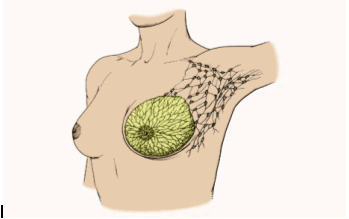
Axillary node dissection:
It is usual practice during breast cancer surgery to remove up to half of the axillary lymph nodes for testing. Testing of the lymph nodes can indicate whether the cancer has spread into the lymphatic system, thus increasing the risk of the cancer spreading to the rest of the body. Axillary node dissection is usually well tolerated but there is a risk that the remaining lymph nodes will not be able to adequately cope with the drainage from the lymphatic vessels in the breast. This can lead to shoulder stiffness, changes in sensation in the area, and a condition known as lymphoedema which is marked by arm swelling.
It is usual practice during breast cancer surgery to remove up to half of the axillary lymph nodes for testing. Testing of the lymph nodes can indicate whether the cancer has spread into the lymphatic system, thus increasing the risk of the cancer spreading to the rest of the body. Axillary node dissection is usually well tolerated but there is a risk that the remaining lymph nodes will not be able to adequately cope with the drainage from the lymphatic vessels in the breast. This can lead to shoulder stiffness, changes in sensation in the area, and a condition known as lymphoedema which is marked by arm swelling.
Sentinel node biopsy:
This type of lymph node biopsy is used in some cases to minimise problems associated with axillary node dissection. During a sentinel node biopsy two special dyes are injected around the breast cancer tumour. One is visible to the naked eye during the biopsy surgery and the other is a weak radioactive substance detectable by either a Gamma camera or a hand-held device like a Geiger counter. The dyes drain through the lymph vessels and into the first node to be involved - the sentinel node. This node is then removed for analysis. If the sentinel node is clear of cancer cells, then it can safely be presumed that the cancer has not spread to the rest of the axillary nodes. If, however, the sentinel node is positive for cancer cells a subsequent procedure to remove the remaining lymph nodes would be needed.
This type of lymph node biopsy is used in some cases to minimise problems associated with axillary node dissection. During a sentinel node biopsy two special dyes are injected around the breast cancer tumour. One is visible to the naked eye during the biopsy surgery and the other is a weak radioactive substance detectable by either a Gamma camera or a hand-held device like a Geiger counter. The dyes drain through the lymph vessels and into the first node to be involved - the sentinel node. This node is then removed for analysis. If the sentinel node is clear of cancer cells, then it can safely be presumed that the cancer has not spread to the rest of the axillary nodes. If, however, the sentinel node is positive for cancer cells a subsequent procedure to remove the remaining lymph nodes would be needed.
Breast reconstruction:
After mastectomy, some women may choose to have the breast reconstructed. This can be done at the time of the mastectomy or at a later date. The surgery is usually performed by a plastic surgeon. The aim of breast reconstruction is to recreate a breast that feels and looks as natural as possible.
After mastectomy, some women may choose to have the breast reconstructed. This can be done at the time of the mastectomy or at a later date. The surgery is usually performed by a plastic surgeon. The aim of breast reconstruction is to recreate a breast that feels and looks as natural as possible.
The majority of breast reconstructions are performed using muscle
and tissue taken from the abdomen in a procedure known as a TRAM flap
procedure. Less commonly, muscle and tissue from the back can also be
used in a procedure called a Latissimus Dorsi flap. Artificial breast
implants can also be used to reconstruct the breast. This is usually
done in conjunction with stretching the skin in order to accommodate the
implant. With TRAM flap or Latissimus Dorsi flap breast reconstruction a
hospital stay of 4-6 days and a recovery period of 4-6 weeks can be
expected.
Occasionally the healthy breast needs to be made smaller to match
the breast that has been reconstructed. This surgery would be performed
at the same time as the breast reconstruction.
For women who do not wish to have breast reconstruction, an
external breast prosthesis can be used. This is a jelly-like
breast-shaped mould that fits into a specially fitted bra and comes in a
variety of shapes and sizes. When worn, the appearance is the same as
that of a normal breast. Wearing the prosthesis also helps to maintain
proper balance and posture. The New Zealand government provides funding
of $600 every four years for the prosthesis and bras.
NON-SURGICAL TREATMENT
Additional treatments are commonly given after surgical removal of a
breast cancer. One or a combination of these treatments may be
recommended. An oncologist (cancer specialist) will be involved in
deciding which treatments will be given.
Radiotherapy:
This uses radiation to destroy any cancer cells that may be left in the breast. It is most commonly used after lumpectomy/partial mastectomy. However, it may be used after mastectomy if there was more than one tumour, the tumour was large, or the tumour was growing close to the chest wall. A course of radiotherapy is usually given over4-6 weeks, consisting of daily treatments from Monday to Friday. Side effects of the treatment include severe tiredness and burns similar to bad sunburn on the treated area.
This uses radiation to destroy any cancer cells that may be left in the breast. It is most commonly used after lumpectomy/partial mastectomy. However, it may be used after mastectomy if there was more than one tumour, the tumour was large, or the tumour was growing close to the chest wall. A course of radiotherapy is usually given over4-6 weeks, consisting of daily treatments from Monday to Friday. Side effects of the treatment include severe tiredness and burns similar to bad sunburn on the treated area.
Chemotherapy:
This may be given if spread of the cancer is suspected or confirmed and is usually given soon after surgery. Chemotherapy medications can be given by tablet or as injections into the blood stream. Usually it is a combination of both. The medications aim to kill off any cancer cells that may be circulating in the body. There are different strengths and combinations of chemotherapy medications, which are given in cycles. Side effects of chemotherapy treatment may include nausea, hair loss, sores in the mouth and diarrhoea.
This may be given if spread of the cancer is suspected or confirmed and is usually given soon after surgery. Chemotherapy medications can be given by tablet or as injections into the blood stream. Usually it is a combination of both. The medications aim to kill off any cancer cells that may be circulating in the body. There are different strengths and combinations of chemotherapy medications, which are given in cycles. Side effects of chemotherapy treatment may include nausea, hair loss, sores in the mouth and diarrhoea.
Hormone therapy:
For cases where the breast cancer is hormone receptor positive, hormone therapy may be prescribed to help prevent recurrence of the breast cancer. These medications work by blocking the hormone receptors on the breast cancer cells, preventing hormones binding to them and stimulating growth. One common example of this type of medication is tamoxifen. This is commonly given for up to five years after diagnosis of breast cancer. Other types of hormone treatments include anastrozole (Arimidex) or letrozole (Letara).
For cases where the breast cancer is hormone receptor positive, hormone therapy may be prescribed to help prevent recurrence of the breast cancer. These medications work by blocking the hormone receptors on the breast cancer cells, preventing hormones binding to them and stimulating growth. One common example of this type of medication is tamoxifen. This is commonly given for up to five years after diagnosis of breast cancer. Other types of hormone treatments include anastrozole (Arimidex) or letrozole (Letara).
Biological therapy:
This type of treatment includes a class of anticancer medications called “monoclonal antibodies”. These medications are formulated to target cancer cells, rather than normal healthy cells. A monoclonal antibody medication used in New Zealand is trastuzumab (Herceptin). This is used to treat women with HER2 positive breast cancer. Trastuzumab works by binding to the HER2 proteins, preventing them from stimulating the cancer cells to grow. It also acts to “flag” the cancer cells to the body, which then stimulates the immune system to destroy the abnormal cells. In New Zealand, trastuzumab is funded for up to 12 months' use in the treatment of women with HER2 positive breast cancer.
This type of treatment includes a class of anticancer medications called “monoclonal antibodies”. These medications are formulated to target cancer cells, rather than normal healthy cells. A monoclonal antibody medication used in New Zealand is trastuzumab (Herceptin). This is used to treat women with HER2 positive breast cancer. Trastuzumab works by binding to the HER2 proteins, preventing them from stimulating the cancer cells to grow. It also acts to “flag” the cancer cells to the body, which then stimulates the immune system to destroy the abnormal cells. In New Zealand, trastuzumab is funded for up to 12 months' use in the treatment of women with HER2 positive breast cancer.
No comments:
Post a Comment